Subscribe to Newsletter
Receive selected insights on neuro-imaging, forensic medicine and expert review.

Traumatic Brain Injury
August 20, 2026
On June 24, 2025, the Radiological Society of North America (RSNA) issued an updated statement on traumatic brain injury (TBI) imaging. It is short, but in a courtroom it carries weight: it defines what mainstream radiology accepts as standard practice for imaging head trauma, and — just as importantly — what it does not yet accept.
TBI is one of the most common neurologic disorders, with falls, motor vehicle collisions, assaults, and sports injuries leading the causes. Because so many of those events end in litigation, the statement is worth reading closely on both sides of a case.
RSNA frames the purpose of TBI imaging around three goals: detecting injuries that require immediate surgical or procedural intervention, detecting injuries that benefit from early medical therapy or close neurologic supervision, and establishing prognosis so that rehabilitation, family counseling, and discharge planning can be tailored to the patient.
That framing matters in a deposition. Imaging obtained in the emergency department was ordered to answer triage questions, not to quantify long-term impairment for a jury. An expert who understands the original clinical purpose of each study can explain why a negative scan does not close the question of injury — and why a positive finding does not automatically explain every reported symptom.
For adults with signs and symptoms of TBI, RSNA confirms that non-contrast head CT is the first line of imaging in the acute phase, and that it can predict mortality and unfavorable outcomes. Non-contrast CT has a high negative predictive value for excluding the need for neurosurgical intervention in mild TBI.
High negative predictive value is not the same as high sensitivity. A normal CT reliably tells the treating team that the patient does not need the operating room tonight; it does not rule out microstructural axonal injury. Conflating the two is one of the most common errors in TBI briefing.
RSNA states that MRI may be indicated in acute TBI when the non-contrast head CT is normal and there are persistent, unexplained neurological findings. MRI has higher sensitivity than CT for detecting axonal injury in mild TBI patients.
At the same time, the statement does not support routine brain MRI for injury detection in acute mild TBI. It reserves MRI for particular circumstances: persistent neurological, cognitive, or behavioral symptoms, and especially new-onset, progressive, or worsening symptoms. In pediatric patients, RSNA is explicit that imaging should not be routinely obtained to diagnose mild TBI.
This is the paragraph that decides admissibility fights. RSNA classifies diffusion tensor imaging, functional MRI, MR spectroscopy, perfusion imaging, PET/SPECT, and magnetoencephalography as investigational in TBI. In RSNA's words, there is insufficient evidence supporting the routine clinical use of these advanced techniques for diagnosis or prognostication of TBI at the individual patient level.
Researchers continue to study whether these techniques can identify injury when conventional CT and MRI are normal, and whether they help with prognostication in patients with persistent symptoms. That research is real and ongoing — but a retained expert who presents an advanced-technique finding as established individual-level proof is exposed on cross-examination, and opposing counsel should know exactly which sentence to read back.
RSNA reiterates that appropriate precautions should always be taken to minimize radiation exposure under the As Low As Reasonably Achievable (ALARA) principle. In cases involving repeat CT imaging — particularly in children — dose stewardship can itself become a standard-of-care issue rather than a footnote.
Our reports cite the RSNA statement where it is directly on point: whether the modality chosen matched the clinical question, whether follow-up imaging was appropriately triggered, and whether an opposing opinion rests on techniques that mainstream radiology still considers investigational.
If your case turns on TBI imaging, an independent review measured against current society statements is the fastest way to learn how strong your imaging evidence really is.
Our content is verified by independent specialists to ensure the highest standards of clinical and forensic accuracy.
| Reviewer | Specialty | Contact Info |
|---|---|---|
| Dr. Saman Hazany, MD, DABR | Neuroradiology | shazany@neuroexpertsgroup.com |
Last reviewed: August 20, 2026
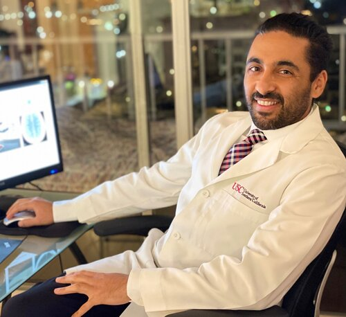
Dr. Saman Hazany is a Harvard-trained Traumatic Brain and Spine Injury Imaging Expert and Chief of Neuroradiology at the Greater Los Angeles VA Healthcare System. He holds academic appointments at major academic centers (UCLA and USC) with a focus on advanced neuro-imaging, including Diffusion Tensor Imaging (DTI), in concussion/TBI. With over 18 years of experience, he is a leading expert witness and educator in neuroradiology.
Contact
Connect with our team for board-certified forensic expertise, rapid report turnaround and testimony that stands up under scrutiny.
Receive selected insights on neuro-imaging, forensic medicine and expert review.
Share your experience with our medical-legal reviews and help us improve our expert resources.
Timely updates from the fields of neuroradiology, neurology and forensic medicine.