Subscribe to Newsletter
Receive selected insights on neuro-imaging, forensic medicine and expert review.

Advanced Imaging
June 24, 2026
In neuro-oncology, every decision matters, and determining whether a brain lesion is aggressive, identifying the true extent of a tumor, or distinguishing tumor recurrence from treatment-related changes can significantly influence patient outcomes. While conventional MRI has long been the cornerstone of brain tumor imaging, it often leaves clinicians with critical unanswered questions, and this is where advanced MRI techniques, along with technologies like DTI neuroimaging , are helping to transform the neuro-oncology landscape.
By moving beyond anatomical images and revealing physiological, metabolic, and cellular information, these technologies are facilitating more precise diagnoses, personalized treatment planning, and improved monitoring of therapeutic response, causing a paradigm shift in how brain tumors are evaluated and managed.
Traditional MRI works incredibly well for showing the size, location, and structural characteristics of brain tumors. Still, it may struggle to differentiate active tumor tissue from inflammation, radiation injury, or post-surgical changes. However, advanced MRI techniques can provide deeper insights into tumor biology, which can help clinicians answer questions such as:
- Has the tumor returned, or is this a treatment effect?
- Which area should be targeted for biopsy or surgery?
In fact, techniques such as diffusion-weighted imaging (DWI), perfusion-weighted imaging (PWI), and MR spectroscopy (MRS) often offer significant advantages in evaluating tumor extent, grading, and treatment response.
Here’s a brief on some of the key advanced MRI techniques that are used in neuro-oncology:
One of the greatest challenges in neuro-oncology is distinguishing true tumor progression from pseudoprogression or radiation necrosis, but on standard MRI, these conditions can appear remarkably similar.
On the other hand, advanced MRI addresses this challenge by examining tissue physiology rather than relying solely on structural changes. For example, perfusion imaging evaluates blood volume within lesions. At the same time, spectroscopy can analyze metabolic signatures that tend to indicate active tumor growth, and this additional layer of information can improve diagnostic confidence while supporting more informed treatment decisions and improving patient outcomes.
The truth about diagnoses in neuro oncology is that the future is multiparametric. The real power of advanced MRI lies in combining multiple techniques into a comprehensive multiparametric assessment. Instead of viewing a tumor from a single perspective, clinicians can evaluate its structure, blood supply, metabolism, and cellular architecture simultaneously.
As imaging technology continues to evolve, advanced MRI is becoming more than a diagnostic tool; it’s emerging as a critical decision-making platform that guides personalized neuro-oncology care seamlessly.
Our content is verified by independent specialists to ensure the highest standards of clinical and forensic accuracy.
Dr. Saman Hazany is a Harvard-trained neuroradiologist and Chief of Neuroradiology at the Greater Los Angeles VA. With over 18 years of experience and faculty roles at UCLA and USC, he specializes in identifying traumatic brain injuries using advanced DTI imaging. As the CEO of Neuro Experts, PC, he is a leading expert witness and educator in the neurological field.
Our content is verified by independent specialists to ensure the highest standards of clinical and forensic accuracy.
| Reviewer | Specialty | Contact Info |
|---|---|---|
| Dr. Saman Hazany, MD, DABR | Neuroradiology | shazany@neuroexpertsgroup.com |
Last reviewed: June 24, 2026
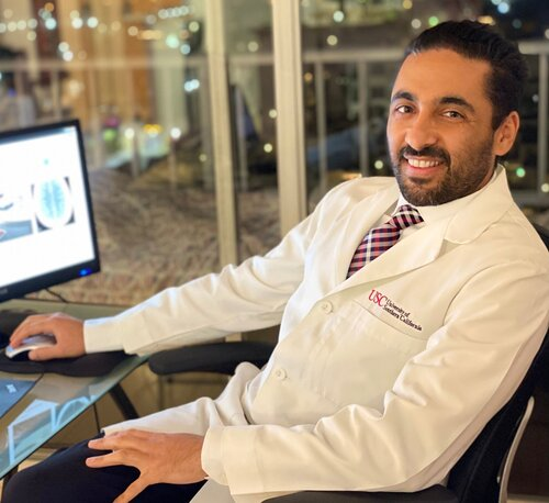
Dr. Saman Hazany is a Harvard-trained Traumatic Brain and Spine Injury Imaging Expert and Chief of Neuroradiology at the Greater Los Angeles VA Healthcare System. He holds academic appointments at major academic centers (UCLA and USC) with a focus on advanced neuro-imaging, including Diffusion Tensor Imaging (DTI), in concussion/TBI. With over 18 years of experience, he is a leading expert witness and educator in neuroradiology.
Contact
Connect with our team for board-certified forensic expertise, rapid report turnaround and testimony that stands up under scrutiny.
Receive selected insights on neuro-imaging, forensic medicine and expert review.
Share your experience with our medical-legal reviews and help us improve our expert resources.
Timely updates from the fields of neuroradiology, neurology and forensic medicine.