Subscribe to Newsletter
Receive selected insights on neuro-imaging, forensic medicine and expert review.

Neuro-Oncology
April 30, 2026
When a workplace injury, a suspected case of medical malpractice, or even a minor accident leads to persistent headaches, memory lapses, or unexpected seizures, the situation can quickly escalate to being more serious than it first appears. What may initially be dismissed as a trauma-related issue can sometimes uncover an underlying condition like a Glioma, which is only visible through detailed imaging! Gliomas are among the most prevalent brain tumors, accounting for roughly 33% of all cases. They develop from glial cells, the supportive cells in the brain that surround and assist neurons, including astrocytes, oligodendrocytes, and ependymal cells. ( Source )
In such cases, timely, unbiased, and accurate diagnosis doesn’t just stand out as a medical priority, but a legal and life-altering choice. Wondering how? Here’s a blog to help you get started!
Medically speaking, Gliomas arise from glial cells, which form the supportive framework of the brain, but every Glioma behaves differently. While some grow slowly and remain localized, others are aggressive and infiltrative, and this variation makes grading and characterization crucial, not just for diagnosis, but also for determining the treatment strategies and predicting outcomes.
Modern imaging techniques like MRI help to evaluate the stance of gliomas, as they help answer critical questions like:
- Has it spread to the surrounding areas?
In fact, MRI sequences, such as T1, T2, FLAIR, and contrast-enhanced scans, provide a layered understanding of the tumor structure, edema, and vascularity. Especially advanced imaging approaches like diffusion-weighted imaging (DWI) and perfusion MRI further refine the picture of gliomas, so they become clearly visible.
Typically, gliomas are classified into four grades by the World Health Organization (WHO), and these grades range from low-grade (I–II) to high-grade (III–IV). Here’s a quick breakdown that breaks down more about the grading before the biopsy:
These are some characteristics that often hint at the tumor’s biological behavior, while guiding clinicians even before histopathological confirmation.
Characterizing gliomas goes way beyond grading, as it involves a comprehensive understanding of the tumor composition, cellular density, and metabolic activity. Advanced tools like MR spectroscopy can detect biochemical changes within the tumor, such as increased choline levels (which indicate the cell membrane turnover) or reduced N-acetylaspartate (which suggests neuronal loss). Diffusion imaging helps assess tumor cellularity, while perfusion imaging tends to evaluate the blood flow, which is often higher in aggressive tumors.
Accurate imaging doesn’t just inform diagnosis; it also shapes the entire treatment pathway, and in some cases, even the legal clarity. For instance:
- In surgical planning, diagnosis helps to determine the resectability and guides neurosurgeons about the glioma.
- In radiation therapy, imaging helps define the target zones, so professionals can spare healthy tissues.
- In monitoring, imaging tracks treatment response and detects recurrence early.
- And, in medico-legal insight, it differentiates pre-existing tumors from trauma-induced symptoms in injury or malpractice claims.
Gliomas are not just identified; they are decoded through imaging. Whether symptoms arise after an injury, a delayed diagnosis, or seemingly out of nowhere, imaging can play a decisive role in uncovering the truth. Thus, if neurological symptoms have emerged after an injury or there are concerns about a missed or delayed diagnosis, expert evaluation is essential.
Connect with Neuro Experts, PC for advanced imaging insights, precise diagnosis, and expert-backed clarity, when it matters the most.
Dr. Saman Hazany is a Harvard-trained neuroradiologist and Chief of Neuroradiology at the Greater Los Angeles VA. With over 18 years of experience and faculty roles at UCLA and USC, he specializes in identifying traumatic brain injuries using advanced DTI imaging. As the CEO of Neuro Experts, PC, he is a leading expert witness and educator in the neurological field.
Our content is verified by independent specialists to ensure the highest standards of clinical and forensic accuracy.
| Reviewer | Specialty | Contact Info |
|---|---|---|
| Dr. Saman Hazany, MD, DABR | Neuroradiology | shazany@neuroexpertsgroup.com |
Last reviewed: April 30, 2026
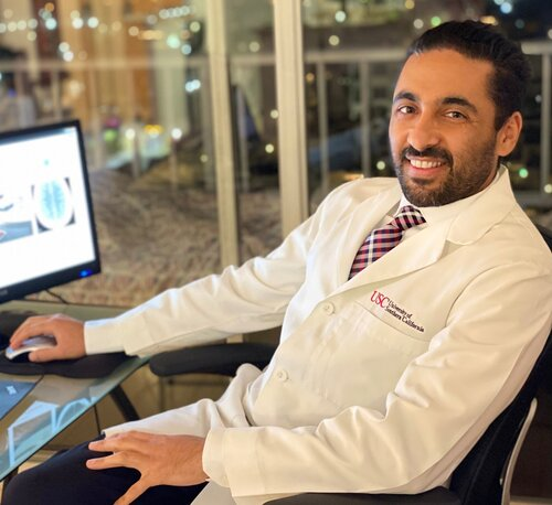
Dr. Saman Hazany is a Harvard-trained Traumatic Brain and Spine Injury Imaging Expert and Chief of Neuroradiology at the Greater Los Angeles VA Healthcare System. He holds academic appointments at major academic centers (UCLA and USC) with a focus on advanced neuro-imaging, including Diffusion Tensor Imaging (DTI), in concussion/TBI. With over 18 years of experience, he is a leading expert witness and educator in neuroradiology.
Contact
Connect with our team for board-certified forensic expertise, rapid report turnaround and testimony that stands up under scrutiny.
Receive selected insights on neuro-imaging, forensic medicine and expert review.
Share your experience with our medical-legal reviews and help us improve our expert resources.
Timely updates from the fields of neuroradiology, neurology and forensic medicine.