Subscribe to Newsletter
Receive selected insights on neuro-imaging, forensic medicine and expert review.

What happens when the cause of death in an investigation case isn’t obvious, and every second of delay means more uncertainty for families, investigators, and courts? This is where an invasive autopsy is done to find out the cause of death. While conventional autopsies have long been the gold standard in postmortem investigations, but it is physically disruptive and emotionally taxing for grieving families.
This is why today, postmortem neuroradiology is used to find deep-seated brain injuries or subtle vascular ruptures without performing any physical dissection. By using advanced imaging procedures like CT and MRI scans, neuroradiology experts can “ look inside” the brain and spinal cord without any incision, and today we are going to talk all about it!
Neuroradiology focuses on imaging the brain, spine, and nervous system, and in postmortem investigations, this helps uncover hidden injuries, diseases, or abnormalities that may explain how a person died. Forensic specialists commonly use the following tools for virtual autopsy or “virtopsy”:
Imagine a suspicious death involving head trauma. A postmortem CT scan can quickly reveal skull fractures or internal bleeding, while an MRI can help to detect soft tissue injuries or signs of stroke. In fact, postmortem CT can show exceptional accuracy in detecting traumatic extra-axial hemorrhage, which makes it incredibly reliable in a forensic diagnosis.
- Traumatic Brain Injuries: Postmortem neuroradiology can help to detect falls, assaults, or accidents that may cause hemorrhages, skull fractures, or diffused axonal injury.
- Stroke or Aneurysm: Sudden deaths from ruptured aneurysms or intracranial bleeds can be spotted through vascular imaging.
- Spinal Cord Injuries: Fatal cervical injuries may not be externally visible, but they can be clearly seen through imaging.
- Foreign Objects: Bullets, shrapnel, or medical devices can also be precisely located through neuroradiology imaging.
Unlike traditional autopsies, neuroradiology offers:
- Better visual evidence for legal proceedings
- Useful in religious or cultural situations where an autopsy is restricted
Especially, postmortem CT is increasingly used worldwide as a supplementary tool, and in some cases, an alternative to full autopsy.
As imaging technology evolves, postmortem neuroradiology will only become more powerful. Besides, when answers matter in high-stakes cases, seeing beneath the surface of autopsies can make all the difference and predict real causes that may have caused the death.
Our content is verified by independent specialists to ensure the highest standards of clinical and forensic accuracy.
| Reviewer | Specialty | Contact Info |
|---|---|---|
| Dr. Saman Hazany, MD, DABR | Neuroradiology | shazany@neuroexpertsgroup.com |
Last reviewed: April 15, 2026
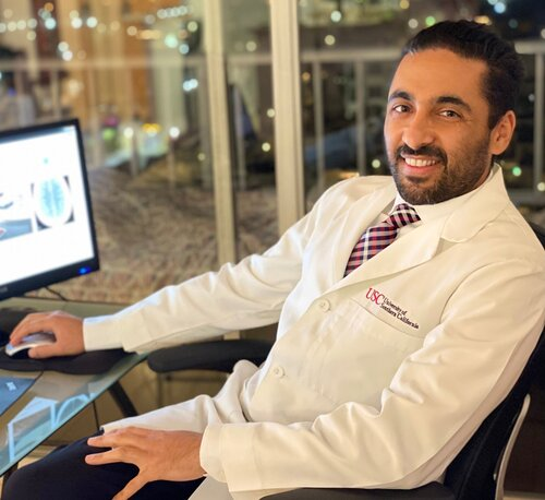
Dr. Saman Hazany is a Harvard-trained Traumatic Brain and Spine Injury Imaging Expert and Chief of Neuroradiology at the Greater Los Angeles VA Healthcare System. He holds academic appointments at major academic centers (UCLA and USC) with a focus on advanced neuro-imaging, including Diffusion Tensor Imaging (DTI), in concussion/TBI. With over 18 years of experience, he is a leading expert witness and educator in neuroradiology.
Contact
Connect with our team for board-certified forensic expertise, rapid report turnaround and testimony that stands up under scrutiny.
Receive selected insights on neuro-imaging, forensic medicine and expert review.
Share your experience with our medical-legal reviews and help us improve our expert resources.
Timely updates from the fields of neuroradiology, neurology and forensic medicine.