Subscribe to Newsletter
Receive selected insights on neuro-imaging, forensic medicine and expert review.

Did you know that Epilepsy affects over 50 million people worldwide? Yet for many individuals, the root cause of their seizures remains elusive. Yes, there’s no denying that EEGs (electroencephalograms) and clinical history provide valuable clues to detect epilepsy. But they often fail to identify the structural abnormalities responsible for seizures. That’s where neuroradiology experts come into the big picture to improve patient outcomes and support clinical research.
(Source: https://www.who.int/news-room/fact-sheets/detail/epilepsy .)
The diagnostic gap in identifying neuroradiology disorders often complicates treatment plans, especially for patients with drug-resistant epilepsy. To bridge this gap, neuroradiology services bring high-resolution imaging and advanced techniques to the forefront of epilepsy evaluation.
Neuroradiology is a subspecialty of radiology that focuses on diagnosing and treating neurological conditions using imaging techniques. It includes modalities like MRI (Magnetic Resonance Imaging), CT (Computed Tomography), PET (Positron Emission Tomography), and functional MRI (fMRI). In diagnosing epilepsy, neuroradiologists collaborate with neurologists, neurosurgeons, and epileptologists to interpret these images and provide insights into the structural and functional state of the brain.
MRI is the gold standard for detecting the structural causes of epilepsy. It can reveal abnormalities such as hippocampal sclerosis, cortical dysplasia, brain tumors, vascular malformations, and previous injuries and infections. High-resolution epilepsy-protocol MRI scans can offer better sensitivity, which helps neuroradiologists detect subtle changes that may be missed on routine imaging.
In cases of temporal lobe epilepsy, the most common focal epilepsy, such as hippocampal atrophy or sclerosis, often appears clearly on MRI. Identifying such lesions can guide decisions on surgical intervention and predict outcomes. For pediatric patients, MRI can also detect congenital malformations like lissencephaly or heterotopia that might indicate seizure activity.
When structural imaging is inconclusive, functional imaging steps in. PET and SPECT scans can identify regions of altered metabolism or blood flow associated with seizure activity. For instance, interictal PET scans using fluorodeoxyglucose (FDG) can highlight hypometabolic zones indicative of epileptogenic foci.
Neuroradiology professionals can provide unbiased results using fMRI and diffusion tensor imaging for pre-surgical planning and clinical research. These tools can help map critical brain functions, such as language, memory, and motor skills, to avoid damaging these areas during the resection of seizure-generating tissue.
For patients who have drug-resistant epilepsy, surgery may be the best chance at seizure control or even remission. Neuroradiology helps evaluate surgical candidacy by locating the focus of the seizure and ruling out multifocal epilepsy. It helps identify eloquent cortex regions to avoid surgery, and 3D reconstructions and advanced imaging sequences further assist in accurate planning as well as intraoperative navigation.
Neuroradiology bridges the gap between symptoms and solutions in epilepsy care. As imaging technology continues to evolve, so will the accuracy and precision of epilepsy evaluation, offering renewed hope for patients living with this complex neurological disorder. For clinicians and patients alike, the integration of neuroradiology into epilepsy management is not only beneficial but also indispensable.
Looking for neuroradiology services? Reach out to our team at Neuro Experts today!
Our content is verified by independent specialists to ensure the highest standards of clinical and forensic accuracy.
| Reviewer | Specialty | Contact Info |
|---|---|---|
| Dr. Saman Hazany, MD, DABR | Neuroradiology | shazany@neuroexpertsgroup.com |
Last reviewed: May 12, 2025
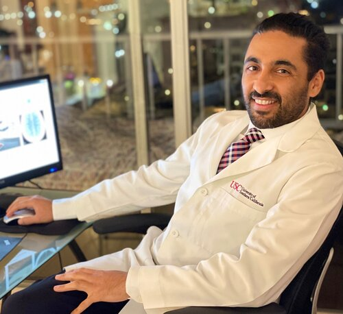
Dr. Saman Hazany is a Harvard-trained Traumatic Brain and Spine Injury Imaging Expert and Chief of Neuroradiology at the Greater Los Angeles VA Healthcare System. He holds academic appointments at major academic centers (UCLA and USC) with a focus on advanced neuro-imaging, including Diffusion Tensor Imaging (DTI), in concussion/TBI. With over 18 years of experience, he is a leading expert witness and educator in neuroradiology.
Contact
Connect with our team for board-certified forensic expertise, rapid report turnaround and testimony that stands up under scrutiny.
Receive selected insights on neuro-imaging, forensic medicine and expert review.
Share your experience with our medical-legal reviews and help us improve our expert resources.
Timely updates from the fields of neuroradiology, neurology and forensic medicine.